
Hormone Types
Introduction
Although hormones work in the same general way, the exact details of how each is created, released, transported, and ultimately exerts its effect vary based on the broader classification of the hormone. In total their are three types of hormones: peptide, steroid, and amino-acid derived. The MCAT tends to test these conceptually and will often require you to spot examples of each hormone based on structure, name, or description.
Peptide Hormones
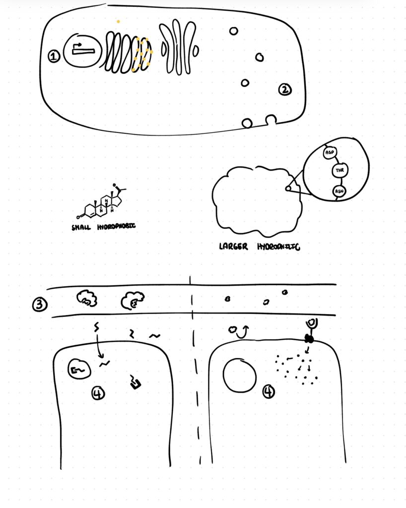
Peptide hormones as their name suggest are strings of amino acids and as with any peptide or protein they are encoded in the DNA and generated through transcription and translation. Ultimately they are packaged up in vesicles and after an incoming signal triggers them they are released into the bloodstream.
Chemically peptide hormones are large and hydrophilic so post release they freely travel dissolved in the aqueous portion of the blood until they reach their target. Since they are polar they cannot cross the hydrophobic membrane of their target cell and must instead bind to surface receptors that kick off secondary messenger cascades within cells.
The MCAT generally will not explicitly tell you that something is a peptide hormone but instead will expect you to know how to spot examples of peptide hormones. While you could memorize a list of every single peptide hormone this is a waste of time as peptide hormones generally share the same naming features, typically they end with -in or hormone. For example, insulin and luteinizing hormone are both peptide hormones. (See table 1 below for more examples)
Content To Passage: Insulin
Instructions: For each paragraph write out a tag and identify any content it is testing then compare your analysis to mine
Phosphorylation of PDX1 causes its translocation to the nucleus where it binds with the A3 element in the insulin promoter where it synergistically works with other coactivators to promote the transcription of preproinsulin, the inactive 110 amino acid long precursor to insulin.
Preproinsulin is translated directly into the rough endoplasmic reticulum (RER), where it undergoes cleavage to remove the signal peptide forming proinsulin. Proinsulin then folds on itself and the ends of the protein called the A chain and B chain are fused together with three disulfide bonds and are subsequently packaged into specialized secretory vesicles. In these vesicles proinsulin is converted to active insulin via proprotein convertases, which cleave out a central portion of proinsulin called the C-peptide.
When blood glucose levels are high, glucose moves into beta-islet pancreatic cells through GLUT2 transporters and through a series of steps triggers calcium influx. The increase in intracellular calcium concentration triggers fusion of the insulin containing vesicles with the cell membrane and release of insulin into the blood stream. Insulin then travels through the blood to its target cells such as a muscle cell and binds to an insulin receptor, a transmembrane receptor tyrosine kinase, and triggers a messenger cascade that ultimately results in the translocation of GLUT4 transporters into the cell membrane allowing glucose to pass into the muscle cell.
View Analysis
Phosphorylation of PDX1 causes its translocation to the nucleus where it binds with the A3 element in the insulin promoter where it synergistically works with other coactivators to promote the transcription of preproinsulin, the inactive 110 amino acid long precursor to insulin.
Tag: Insulin transcription
- PDX1 is a transcription factor and this is a textbook example after receiving a signal it moves to the nucleus and binds with the DNA (insulin promoter) to start transcription
- Insulin is a peptide hormone the -in suffix is a dead give away that this is a protein
- Preproinsulin the pre tells you this protein has a signal sequence which would be needed for a secreted peptide hormone and the pro lets you know this has an inactive form prior to forming active insulin
- The 110 amino acids could be used in a question asking about the Dalton weight or number of nucleotides in preproinsulin
Preproinsulin is translated directly into the rough endoplasmic reticulum (RER), where it undergoes cleavage to remove the signal peptide forming proinsulin. Proinsulin then folds on itself and the ends of the protein called the A chain and B chain are fused together with three disulfide bonds and are subsequently packaged into specialized secretory vesicles. In these vesicles proinsulin is converted to active insulin via proprotein convertases, which cleave out a central portion of proinsulin called the C-peptide.
Tag: Preproinsulin processing
- Translation directly into the RER suggests a protein is entering the secretory pathway or being trafficked to a specific organelle or the plasma membrane this is exactly what you would expect for a peptide hormone like insulin
- Proinsulin folding in on itself and joining together by disulfide bonds is an example of tertiary structure
- That fact that disulfide bonds can form suggests proinsulin is in an oxidizing environment which is typical of the RER, Golgi, and secretory vesicles
- Proprotein convertases are not something you should know outright but you should know they are enzymes from the -ase suffix and deduce they are hydrolases because they cleave proteins which is most commonly acomplished via hydrolysis
When blood glucose levels are high, glucose moves into beta-islet pancreatic cells through GLUT3 transporters and through a series of steps triggers calcium influx. The increase in intracellular calcium concentration triggers fusion of the insulin containing vesicles with the cell membrane and release of insulin into the blood stream. Insulin then travels through the blood to its target cells such as a muscle cell and binds to an insulin receptor, a transmembrane receptor tyrosine kinase, and triggers a messenger cascade that ultimately results in the translocation of GLUT4 transporters into the cell membrane allowing glucose to pass into the muscle cell.
Tag: Calcium releases insulin
- Glucose as a polar molecule can’t freely cross the membrane it must use a channel or transporter here that is GLUT3
- Notice how similar hormone release is to neurotransmitter release they work on the same system and are triggered by the same thing calcium influx
- From release of insulin to insertion of GLUT4 transporters the rest of the passage is simply an example of how peptide hormones exert their effects.
- Travel freely in the blood: Insulin then travels through the blood
- Bind cell surface receptors: binds to an insulin receptor, a transmembrane receptor tyrosine kinase,
- Act through messenger cascades: triggers a messenger cascade that ultimately results in the translocation of GLUT4
Steroid Hormones
Steroid hormones are cholesterol derivatives and contain the characteristic four ring steroid structure.
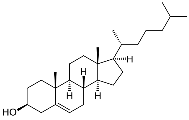
They are created in the smooth endoplasmic reticulum of cells and as smaller, hydrophobic molecules that can easily cross cell membranes they freely diffuse from the cells where they are created into the blood. Unlike their polar, peptide counterparts they require carrier proteins, primarily albumin, to travel through the blood to their targets.
Once at their target cells they diffuse across their plasma membrane and into the cells where they bind cytosolic and nuclear receptors to exert their effects.
Again the MCAT will expect you to be able to spot steroid hormones examples, which you can do by looking out for -sterone, -str-, and -ol in their names. For example testosterone and estriol.
Content To Passage: Aldosterone
Aldosterone is a hormone that regulates systemic blood pressure by promoting sodium and water reabsorption in the distal nephron. Its secretion is controlled primarily by angiotensin II and serum potassium, both of which act on the zona glomerulosa, the outermost zone of the adrenal cortex. Aldosterone biosynthesis is governed at the transcriptional level by aldosterone synthase, encoded by CYP11B2, whose expression is restricted to the zona glomerulosa under normal physiological conditions.
Adapted from: Suzuki D, Saito-Hakoda A, Ito R, Shimizu K, Parvin R, Shimada H, et al. (2017) Suppressive effects of RXR agonist PA024 on adrenal CYP11B2 expression, aldosterone secretion and blood pressure. PLoS ONE
View Analysis
Aldosterone is a hormone that regulates systemic blood pressure by promoting sodium and water reabsorption in the distal nephron. Its secretion is controlled primarily by angiotensin II and serum potassium, both of which act on the zona glomerulosa, the outermost zone of the adrenal cortex. Aldosterone biosynthesis is governed at the transcriptional level by aldosterone synthase, encoded by CYP11B2, whose expression is restricted to the zona glomerulosa under normal physiological conditions.
Tag: Aldosterone secretion and synthesis
- Aldosterone the sterone in the name tells you this is a steroid hormone
- Remember water follows sodium so its no surprise to see water reabsorption tied to sodium reabsorption
- If you ever see angiotensin II you should think of the renin-angiotensin-aldosterone system
- CYP prefix (CYP11B2) tells you this is a cytochrome p450 enzyme
- Gene expression restricted to a particular cell type means the transcription in these cells not the DNA is different. The MCAT loves to trip students up with this remember all of the somatic cells in the body have the same DNA but express that DNA very differently
Explain The Image
Below is a diagram summarizing how both peptide and steroid hormones work. Label as much of the diagram as possible and explain what is happening at each numbered step.
Amino Acid Derivative Hormones
Amino acid derivative hormones are the smallest of the bunch and are created from single amino acids. Their properties and mechanisms of action differ from hormone to hormone and have. You’ll learn these details later when individuals hormones are explored in more depth.
As with all the other hormones types you need to be able to spot these by their -ine suffix. For example dopamine and L-thyroxine.
Hormone Name Summary
| Peptide Hormones | Steroid Hormones | Amino Acid Derivative |
| Oxytocin | Progesterone | Dopamine |
| Somatostatin | Estrogen | Triiodothyrine |
| Calcitonin | Aldosterone | L-thyroxine |
| Growth Hormone | Estradiol | Norepinephrine |
| Follicule Stimulating Hormone | Cortisol | Epinephrine |